The Language of Cells: How Communication Differs in Healthy and Tumour Tissue
What changes when normal pancreatic tissue becomes malignant?
By comparing tumour and adjacent normal tissue, we examined how communication between immune, stromal and other cell populations is rewired in pancreatic cancer.
Pancreatic ductal adenocarcinoma (PDAC) is characterized by a particularly complex tumour microenvironment (TME), where malignant cells coexist with immune cells, fibroblasts, endothelial cells, and other cellular populations. In order to better understand this microenvironment, we need to profile which cell types are present and map how those cell types communicate with one another.
Cell-cell communication (CCC) is fundamental to tissue organization and function. Cells continuously exchange molecular signals that regulate processes such as proliferation, differentiation, immune activity, tissue maintenance, and responses to environmental change. In cancer, these communication networks can be extensively altered, contributing to disease progression and shaping the behaviour of both malignant and non-malignant cells within the tumour.
Within CancerScan, Neovivum Technologies is using cell-cell communication analysis to identify recurring patterns in how signalling networks change in cancer. These patterns feed into Neovivum’s work on tumour “proto-grammar,” which aims to define the transformation rules governing how cellular communication networks are reorganized during tumour development.
For our analysis, we used CellChat v2, a computational framework for inferring cell-cell communication from single-cell RNA-sequencing (scRNA-seq) data, to compare the signalling networks in the tumour tissue with signalling networks in the adjacent normal tissue of the pancreas. The results identified specific cell populations and signalling pathways whose communication patterns are altered the most in the TME, offering a systems-level view of how PDAC reshapes intercellular signalling.
Data processing and clustering
We used the GSE212966 dataset, that contains 69,105 cells from 6 patients. Each patient is represented by two samples: one taken from the PDAC tumour and another from adjacent-normal pancreas tissue. Standard quality-control measures were used to remove low-quality cells and likely doublets, reducing the dataset to 57,232 cells. One normal sample (ADJ3) that showed substantially poorer quality than the others was excluded, leaving five normal biological replicates.
Cell-type identification
The 17 clusters were then assigned biological cell identities using a combination of automated classification and marker-gene analysis. CellTypist identified several immune-cell populations, while additional marker-gene analysis was used for cell types that the immune-focused reference could not classify. This combined approach identified 12 cell types, including T cells, macrophages, fibroblasts, NK (natural killer) cells, endothelial cells, ductal/epithelial cells, and several other immune and pancreatic cell populations.
Cell-cell communication analysis
CellChat was then used to compare communication networks between tumour and adjacent normal tissue. The analysis examined which cell types were sending and receiving signals, how strongly they interacted, and which signalling pathways differed between the two conditions. Plasma cells were excluded from this stage because only five were present in tumour tissue, while NK cells were retained despite their low number because their depletion was considered biologically important.
Key Findings
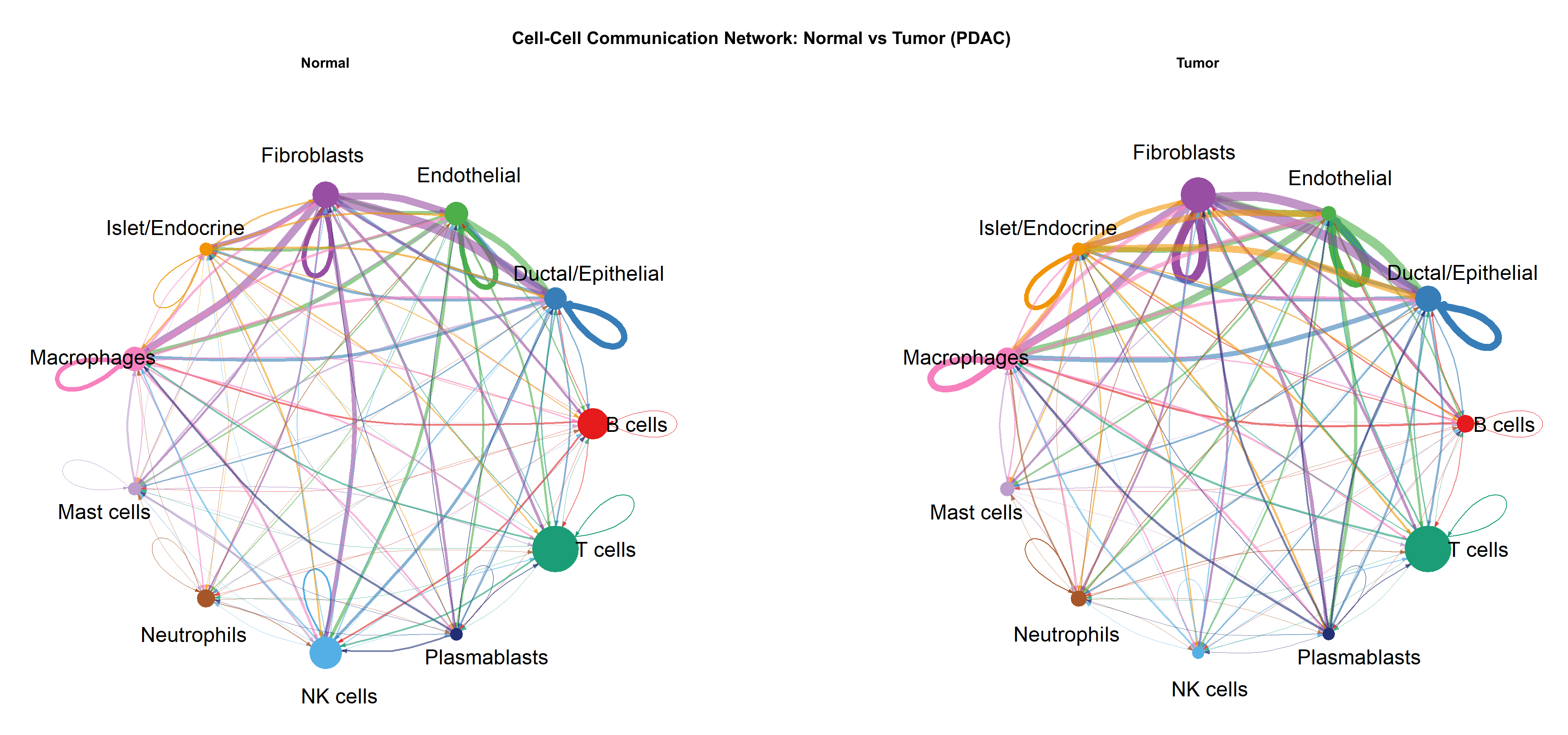
NK cells underwent the single most dramatic shift with a near-total collapse in cell numbers (N3,213 🡪 T71) and a profound change in their network role, moving from the highest incoming-signal cell type in normal tissue to the lowest in tumour. This is consistent with active immune evasion.
Fibroblasts showed the opposite. They expanded more than threefold in tumour tissue (N2,304 🡪 T7,664 cells) and emerged as the loudest sender of signals, especially through COLLAGEN/PERIOSTIN pathways, which is consistent with PDAC’s characteristic desmoplastic stroma.
Macrophages (TAMs) roughly doubled in number and became the network’s leading incoming-signal hub in tumour tissue, consistent with increased recruitment and altered activity of tumour-associated macrophages.
Endothelial cells decreased in number but increased in signalling activity per cell, suggesting an activated angiogenic phenotype rather than passive vessel expansion, which is consistent with PDAC’s hypovascular but angiogenically active tumour biology.
Plasma cells were nearly eliminated in tumour (N2,029 → T5 cells), a finding significant enough to require their exclusion from formal CellChat analysis, but notable in its own right as further evidence of humoral immune suppression.
Interpreting the Results
CellChat provides an inferred view of cell-cell communication based on gene-expression patterns, so the results should be understood as a framework for identifying biologically meaningful changes rather than direct measurement of signalling events. The NK-cell finding is based on a relatively small tumour population of 71 cells, and one adjacent-normal sample was excluded during quality control, leaving five normal biological replicates. These factors should be considered when interpreting individual results, but they do not diminish the broader pattern observed across the analysis: a substantial reorganization of the PDAC microenvironment, marked by the loss of immune-cell signalling and increased stromal and tumour-associated communication.
Conclusion
This analysis adds a quantitative, systems-level view of how that environment is organized, showing which cell populations become more or less prominent, how their signalling roles change, and which pathways are most affected. Rather than describing the tumour microenvironment only by its cellular composition, the CellChat analysis reveals how interactions between those cells are reorganized in PDAC. This provides a clearer basis for identifying signalling relationships and cellular populations that may warrant further experimental investigation.
Note: This article is based on the analysis of publicly available single-cell RNA-sequencing data (GSE212966) using CellChat v2.
References
[1] Jin, S., Guerrero-Juarez, C.F., Zhang, L., Chang, I., Ramos, R., Kuan, C.-H., Myung, P., Plikus, M.V. and Nie, Q. (2021). Inference and analysis of cell-cell communication using CellChat. Nature Communications, 12(1), p.1088.
[2] Gene Expression Omnibus (2023). GSE212966 — Single-cell RNA sequencing of pancreatic ductal adenocarcinoma and adjacent normal tissue. National Center for Biotechnology Information.
Links
https://github.com/jinworks/CellChat
https://www.ncbi.nlm.nih.gov/geo/query/acc.cgi?acc=GSE212966
Keywords
Pancreatic ductal adenocarcinoma, CellChat, single-cell RNA sequencing, tumour microenvironment, cell-cell communication, immune evasion, cancer-associated fibroblasts, tumour-associated macrophages, natural killer cells