Tumor microenvironment (TME) sustains and shapes cancer cell activity
Since the first description of cancer hallmarks by Hanahan and Weinberg 25 years ago (Hanahan and Weinberg, 2000). A striking number of cancer-enabling processes have been identified within the tumor microenvironment (TME). TME is nowadays widely accepted as an integral element in cancer.
The tumor microenvironment is the physical and biological niche supporting cancer cell growth. It comprises the environmental factors found in tumors such as hypoxia, low pH or high extracellular matrix (ECM) content in comparison to the surrounding healthy tissue, along with a particular cell composition e.g. cancer associated fibroblasts, immune infiltrates or endothelial cells among others. All these elements have a direct influence in cancer cell activities and the way it responds to therapy.
Tumor and tumor microenvironment
The way in which tumors are perceived have drifted over the years from a rather simple vision, in which a group of cells become malignant by the accumulation of somatic mutations, to a much more complex and integrative view. Tumors are biological multicellular systems, exquisite in their complexity, heterogeneity, and adaptability (Dagogo-Jack and Shaw, 2018). They are intricately embedded and dependent on the body but possess the ability to adapt to environmental stimuli and evolve. Such plasticity does not solely affect cancer cells but leads to significant changes in healthy cells. For instance, under low oxygen levels, tumor cells facilitate angiogenesis (Harris, 2002). During the metastatic cascade, cancer cells stimulate resident cells supporting local invasion and the distal preconditioning of the metastatic niche (Fares et al., 2020). Additionally, under chemotherapeutic pressure, cancer cells may enter a quiescent drug-resistant state influenced by the tumor microenvironment (TME) (Chen et al., 2021). In the end, this effect can lead to higher chances of tumor relapse (Meads, Gatenby and Dalton, 2009).
Tumor microenvironment in pancreatic cancer
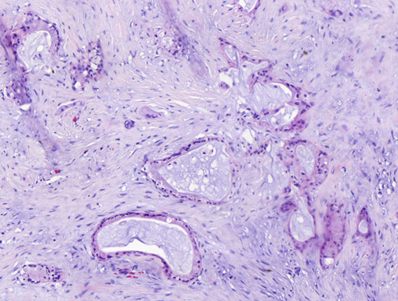
In particular, pancreatic cancer is characterized by highly desmoplastic tumors, in which the TME confers a dense and strong physical barrier preventing the access of anticancer treatments. Within, a spectrum of cell types co-exists, providing cancer cells with a notorious cancer cell plasticity and drug resistance capabilities. For instance, pancreatic cancer dense stroma is the result of cancer-associated fibroblasts (CAFs) activity, whose collagen secretion contributes to the formation of tight extracellular matrices (ECM) (Truong and Pauklin, 2021). CAFs and immune infiltrates have also been shown to modulate cancer cell phenotypes, supporting cancer cell adaptation, by paracrine signalling. In addition, coordinated action of Schwann cells and ECM remodelling capacities from CAFs have been proven to assist cancer cells during perineural invasion processes (Xue et al., 2023).
Tumor and tumor microenvironment cross-talk
This adaptive response significantly impacts disease progression, treatment resistance, and recurrence, and can only be understood in the framework of continuous interplay between tumor cells and their multicellular environment (Ramón y Cajal et al., 2017; Celià-Terrassa and Kang, 2018) . One emerging actor in the two-way communication between TME and cancer cells are extracellular vesicles. EVs are membrane bound vesicles secreted by most cell types that contain a biological cargo (nucleic acids, proteins, metabolites) and are widely involved in intercellular signaling. In cancer, EVs have been shown to impact tumor cells and their microenvironment by promoting epithelial mesenchymal transition (EMT) (Galindo-Hernandez et al., 2015), conferring drug resistance (Boelens et al., 2014; Binenbaum et al., 2018), transforming stroma cells such as mesenchymal stem cells (Chowdhury et al., 2015), fibroblasts (Fang et al., 2018) and endothelial cells (Tang et al., 2018) towards phenotypes prone to foster tumoral growth, in both consolidated tumors and pre-metastatic niches (PMN), or exerting immunosuppressive activities on tumor infiltrating immune cells (Wang et al., 2018; Biswas et al., 2019).
It is clear then, that tumor progression and response to treatment is the result of a multifactorial system comprising i) cancer cells ii) their physical and cellular surroundings and iii) the system capacity to adapt, largely influenced by heterotypic intercellular communication avenues.
In this scenario, CancerScan aims at identifying the major contributors associated to TME by combining a multimodal study that integrates multi-omics data (proteomics, transcriptomics) from clinically validated tumor models and image analysis from clinical biopsies. This will allow us to map the influence of the TME on the efficacy of chemotherapeutic treatments and guide oncologist decision towards personalized therapeutic schemes.
References
Binenbaum, Y. et al. (2018) “Transfer of miRNA in Macrophage-Derived Exosomes Induces Drug Resistance in Pancreatic Adenocarcinoma.,” Cancer research, 78(18), pp. 5287–5299. Available at: https://doi.org/10.1158/0008-5472.CAN-18-0124.
Biswas, S. et al. (2019) “Exosomes Produced by Mesenchymal Stem Cells Drive Differentiation of Myeloid Cells into Immunosuppressive M2-Polarized Macrophages in Breast Cancer,” The Journal of Immunology, 203(12), pp. 3447–3460. Available at: https://doi.org/10.4049/jimmunol.1900692.
Boelens, M.C. et al. (2014) “Exosome Transfer from Stromal to Breast Cancer Cells Regulates Therapy Resistance Pathways,” Cell, 159(3), pp. 499–513. Available at: https://doi.org/10.1016/j.cell.2014.09.051.
Celià-Terrassa, T. and Kang, Y. (2018) “Metastatic niche functions and therapeutic opportunities,” Nature Cell Biology, 20(8), pp. 868–877. Available at: https://doi.org/10.1038/s41556-018-0145-9.
Chen, K. et al. (2021) “The metabolic flexibility of quiescent CSC: implications for chemotherapy resistance,” Cell Death & Disease, 12(9), p. 835. Available at: https://doi.org/10.1038/s41419-021-04116-6.
Chowdhury, R. et al. (2015) “Cancer exosomes trigger mesenchymal stem cell differentiation into pro-angiogenic and pro-invasive myofibroblasts,” Oncotarget, 6(2), pp. 715–731. Available at: https://doi.org/10.18632/oncotarget.2711.
Dagogo-Jack, I. and Shaw, A.T. (2018) “Tumour heterogeneity and resistance to cancer therapies,” Nature Reviews Clinical Oncology, 15(2), pp. 81–94. Available at: https://doi.org/10.1038/nrclinonc.2017.166.
Fang, T. et al. (2018) “Tumor-derived exosomal miR-1247-3p induces cancer-associated fibroblast activation to foster lung metastasis of liver cancer.,” Nature communications, 9(1), p. 191. Available at: https://doi.org/10.1038/s41467-017-02583-0.
Fares, J. et al. (2020) “Molecular principles of metastasis: a hallmark of cancer revisited,” Signal Transduction and Targeted Therapy, 5(1), p. 28. Available at: https://doi.org/10.1038/s41392-020-0134-x.
Galindo-Hernandez, O. et al. (2015) “Extracellular vesicles from women with breast cancer promote an epithelial-mesenchymal transition-like process in mammary epithelial cells MCF10A,” Tumor Biology, 36(12), pp. 9649–9659. Available at: https://doi.org/10.1007/s13277-015-3711-9.
Hanahan, D. and Weinberg, R.A. (2000) “The Hallmarks of Cancer,” Cell, 100(1), pp. 57–70. Available at: https://doi.org/10.1016/S0092-8674(00)81683-9.
Harris, A.L. (2002) “Hypoxia — a key regulatory factor in tumour growth,” Nature Reviews Cancer, 2(1), pp. 38–47. Available at: https://doi.org/10.1038/nrc704.
Meads, M.B., Gatenby, R.A. and Dalton, W.S. (2009) “Environment-mediated drug resistance: a major contributor to minimal residual disease,” Nature Reviews Cancer, 9(9), pp. 665–674. Available at: https://doi.org/10.1038/nrc2714.
Ramón y Cajal, S. et al. (2017) “Cancer as an ecomolecular disease and a neoplastic consortium,” Biochimica et Biophysica Acta (BBA) – Reviews on Cancer, 1868(2), pp. 484–499. Available at: https://doi.org/10.1016/j.bbcan.2017.09.004.
Tang, M.K.S. et al. (2018) “Soluble E-cadherin promotes tumor angiogenesis and localizes to exosome surface,” Nature Communications, 9(1), p. 2270. Available at: https://doi.org/10.1038/s41467-018-04695-7.
Truong, L.H. and Pauklin, S. (2021) “Pancreatic cancer microenvironment and cellular composition: Current understandings and therapeutic approaches,” Cancers. Available at: https://doi.org/10.3390/cancers13195028.
Wang, X. et al. (2018) “Hypoxic Tumor-Derived Exosomal miR-301a Mediates M2 Macrophage Polarization via PTEN/PI3Kγ to Promote Pancreatic Cancer Metastasis,” Cancer Research, 78(16), pp. 4586–4598. Available at: https://doi.org/10.1158/0008-5472.CAN-17-3841.
Xue, M. et al. (2023) “Schwann cells regulate tumor cells and cancer-associated fibroblasts in the pancreatic ductal adenocarcinoma microenvironment,” Nature Communications, 14(1), p. 4600. Available at: https://doi.org/10.1038/s41467-023-40314-w.
Keywords
Pancreatic Cancer, Tumor Microenvironment (TME), Cell Communication, Drug Resistance, Metastasis