Making tumour communication visible: tracking extracellular vesicles in 3D cancer models
Tumours are complex ecosystems in which cancer cells constantly interact with the surrounding microenvironment. These interactions can influence tumour growth, progression and response to treatment. Among the mechanisms that enable this communication are extracellular vesicles (EVs), small particles released by cells that carry biological information to other cells.
One of the major challenges in studying EVs is understanding where they come from. In a tumour, cancer cells, fibroblasts, endothelial cells and other stromal populations can all release EVs. When these vesicles are analysed together, it is difficult to determine which cell populations are responsible for the signals being observed.
Within CancerScan, VHIR researchers are addressing this challenge by developing a strategy to track and quantify EV populations according to their cellular origin in multicellular tumour spheroids.
Following EVs within a tumour-like environment
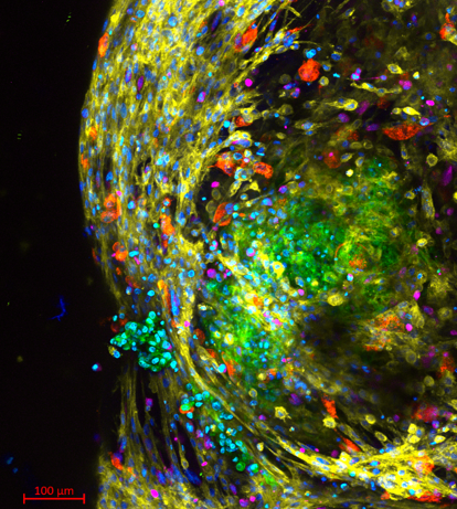
To investigate EV-mediated communication in a more realistic setting, we have established multicellular spheroids combining pancreatic cancer cells with key stromal populations, including cancer-associated fibroblasts, endothelial cells and pancreatic stellate cells. Each cell population can be distinguished through a specific EV label, allowing vesicles released by different cellular sources to be tracked when the cells grow together within the same three-dimensional model.
Imaging studies have confirmed the organisation of the different cell populations within the spheroids, providing a controlled model in which cancer–stroma interactions and EV-mediated communication can be studied simultaneously.
From detecting EVs to understanding their origin
The labelled EVs can be detected and quantified in the material released by the spheroids, allowing researchers to distinguish different EV populations according to their cellular source.
This is particularly important because the total number of EVs released by a tumour model does not necessarily reveal which cells are driving the communication. Being able to resolve EV populations by their origin provides an additional layer of information and opens the possibility of studying how communication between different tumour and stromal populations changes over time or in response to treatment.
The approach also allows selected EV populations to be isolated for further molecular analysis. This will make it possible to investigate not only which cells are releasing EVs, but also what biological information those vesicles carry
Understanding the effects of cancer treatment
A significant use for these models within the CancerScan project is to compare EV-mediated communication in untreated and treated tumour spheroids. Cancer treatment can alter the behaviour of both malignant and stromal cells. These changes may also affect the EVs they release, potentially modifying communication within the tumour microenvironment.
By tracking EV populations from different cellular sources, we aim to determine how treatment reshapes this communication landscape and whether particular EV populations become more prominent under therapeutic conditions.
Building a map of tumour communication
The next phase of the work will combine EV tracking with proteomic analysis to characterise the molecular cargo of selected EV populations. Together, these data will provide three complementary pieces of information: which cells are communicating, which EV populations are involved, and what molecular information they carry.
The resulting datasets will contribute to the broader CancerScan effort to reconstruct communication networks within the tumour microenvironment and integrate them into computational models and knowledge-based frameworks.
By making the origin of EV-mediated signals visible, this approach provides a new way to study the dynamic communication between cancer and stromal cells, and to understand how these interactions change during treatment.
Keywords
Extracellular Vesicle, Tumour Microenvironment, Pancreatic Cancer, Multicellular Models, Cancer Communication combination